#premed

Fall schedule looking rough. My preliminary idea is to take 3 classes, which would make my week look like this:
Monday: work until 11, gen chem on the other campus 30 minutes away 2-4
Tuesday: microbio at the close campus from 8:30-9:30, followed by lab from 10-1, then physiology from 2-4 and lab from 4-7.
Wednesday: work until 11, chem on the far campus from 2-4 followed by lab until 7
Thursday: microbio and lab from 8:30-1, physiology from 2-4
Friday: work
Saturday: off/study/sleep
Sunday: work
I’ve got to get another semester of gen bio (that will be easy) a year of chem, a year of physics (technically I have credit for this and chem but I sucked at it so I should probably take it again), biochem and a year of o chem (have to take chem first). All with labs. And studying for the MCAT. And getting experience/science electives where I can. I want to take phlebotomy but it requires an externship and I have to find out what that entails time-wise because it’s not REQUIRED but it would be super useful and was suggested by current med students to take premed if possible. I ALREADY HAVE A BACHELOR’S DEGREE. I’m gonna be in college for literally 15 years. I graduated in 2010. Fml.
I can’t not work because even as it is I’m not making enough to save anything. I don’t know what I’m gonna do about that. I’m gonna try to get a job over the summer and save everything from it. My car gets better gas mileage now so that’s an extra 100 a month but commuting to the far campus will eat that up.
I hope I won’t have to go back to PT cuz I won’t have any time to go! Therapy’s gonna have to wait but I won’t have time to be crazy anyway. I’ll have to cut down on meetings too but I also won’t have any money to buy booze.


8.01.2019
«Let the chemistry game begin and may the odds be ever in your favor!»
P.s. my mother accepted my decision, yahooo, so LETS WORK AND ETC, FIGHTING
P.s.s. OMG, chemistry is so interesting, hard, but really interesting, I am so sad that my teachers at school really sucked in explanations and etc, that made me even hate this subject (
Post link
Opportunityfor Girls Interested in STEAM!
Hi everyone! I’m a writer for a nonprofit organization called Girl Genius (@girlgeniusmag on insta!). GG strives to create an inclusive community where girls of all backgrounds in STEAM (non-binary and trans-inclusive) are given a voice to empower the next generation of female change makers! I wanted to share an upcoming event with you all!
OnJuly 5, 2020, Girl Genius is launching their first online conference (it’s completely free to participate!). There will be many panels and workshops! There are also prizes (ex. Kankens, bullet journal supplies, speakers, hydroflasks) being raffled off!
The panels and workshops include:
• Women in STEAM
• College Application Panel
• Pre-med Panel
• Getting Research Internahips In High School 101 Workshop
• Intro to Animaltion Workshop
• Breaking into Tech Workshop (Vercel x Girl Genius)
• AR/VR Workshop
You do not have to stay for the entire conference, you can just attend the panels/workshops you are interested in! Once you register, you will get an email a day before the event begins!
Here is some more info on some of the panels! ↓


Here is the link to register!
*the last question asks if you found out about this conference through someone’s social media! I would appreciate it if you would put down my name (Bidhi) for that question! Thank you!! :)
Hope to see you all there! Feel free to message me with any questions!

My second round of midterms is already just six days away! Time to get serious and review some lectures.
Post link


Snapshots of a Past Study Sesh feat. My weapon of choice, the song of the hour, and the finished product~
Post link

I’m lovingggg this Hormones and Behavior class!! It’s so interesting to learn about how our bodies work and what happens when things don’t go perfectly.
Post link


Esoph-egg-us
✅Non-keratinized stratified squamous epithelium
✅Circumscribing muscularis mucosae
✅Submucosal glands
✅Dual layered muscularis externa
It’s just not Spring until you have painted your very own Esoph-egg-us
So get cracking and I don’t want to hear any eggscuses
#histology #science #pathology #pathologists #anatomy #autopsy #eggs #easter #spring #biology #esophagus #digestive #premed #meded #nurse #nursing #medschool #medstudent #medicine #education #vetscience #vetschool #dentistry #histotechnology #histologica #histotech #histo #pathArt #sciArt #ihearthisto

Grumpy Cord
A transverse slice through a spinal cord that looks like Grumpy Cat.
Being the center of the nervous system, transmitting neural signals between the brain and the entire body and controlling independent neural reflexes…is just so awful .
The pale central face is the ‘grey matter’ and is home to the many neuron cell bodies that run through the spinal cord. The surrounding darker region is composed of the axons of these neurons as the exit, enter, ascend and descend through the spinal cord on their way to innervate muscles, or returning information about pain back from the skin, or relaying information about body position back to the brain.
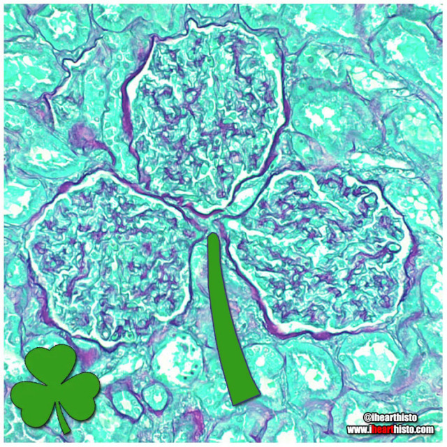
☘️Shamrock Gloms ☘️
For each petal on the shamrock,
This brings a wish your way.
Good health. Good luck. Happiness.
For today and every day.
Happy St. Patrick’s Day!
Three renal corpuscles (glomerulus + their surrounding Bowman’s capsules) floating in a sea of distal and proximal convoluted tubules within the cortex of the kidney.
These three small structures are knotted balls of capillaries (glomeruli) surrounded by a specialized epithelium (Bowman’s capsule) that is composed of cells called podocytes. These cells have tiny interlocking legs that form a small slit between them.
This structural organization is responsible for filtering your blood to produce a fluid that then travels within tubes continuous with the Bowman’s capsule called nephrons. In these nephrons the tubular fluid is modified by reabsorbing and secreting ions and conserving water to produce urine for excretion.

⚡Lord Voldermis⚡
A biopsy of a region of skin-that-shall-not-be-named (dermis/hypodermis junction shhh), complete with nerves, vessels, sweat glands and hair follicles.
by @nejiby

You can find LOVE in the strangest of places (2022 edition)
By row starting top left:
1. in a skin cylindroma
2. in an hepatic ductule
3. in a pancreas
4. in a warty penile growth
5. in a mucus-y colon
6. in a region of hypodermis
7. in a secondary oocyte
8. in a chondrosarcoma
9. in a small artery
Happy Valentine’s Day
Tag a friend with the histo heart you want to share with them and spread the love!
Images by:
@ihearthisto [1-4, 5-9]
@donna.horncastle [5]
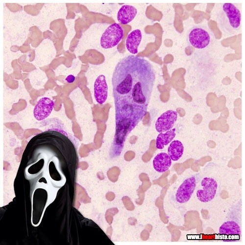
Ghostface Killer is back!
Only this time he’s hiding in a rectum!
Scaring while you’re learning!
A rectal scrape is performed (more often in veterinary med) to obtain a sample of the epithelial cells lining the rectum. These are observed in the microscope for abnormal changes or for microbes and their eggs/larvae that might be present in the digestive tract.
This particular scrape contains an epithelial cell that appears to be bi-nucleate and is surrounded by red blood cells.

Cookie Monster Ovary
Today’s ihearthisto is brought to you by the letter O for Oocyte!
This is image shows a high magnification view of a slice through an ovary.
You are looking at four tiny primordial follicles (the cookies above Cookie Monster’s head) located in the outer cortex of the ovary. Each of these follicles contains a single dormant, immature egg (a primary oocyte) that is halted in the the first phase of meiosis (prophase I). Eventually these follicles will be recruited to enter the maturation cycle that, against all the odds, could see them develop into an embryo.
Cookie monster himself is a larger multilaminar primary follicle (the cookie in his mouth is the nucleus of the oocyte within the follicle). This type of follicle has already been recruited and it’s follicular cells are dividing and differentiating. The oocyte within it though is still halted in prophase I of meiosis.
An oocyte will only complete meiosis if it is ovulated and fertilized by a spermatozoon (a sperm cell).
Post link
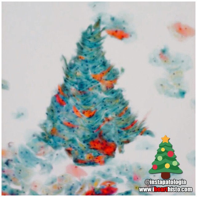
❄️Pappy Holidays❄️
There’s nothing quite like a Pap smear Christmas tree to rock around this happy holiday season!
by the awesome @instapatologia [Insta]

Have a Howelly-Jolly Christmas
A festive finding in the blood of an asplenic patient
A Howell–Jolly body is a cytopathological finding whereby small remnants of nuclear DNA are present in normally anuclear circulating erythrocytes.
During development in the bone marrow, late orthochromatophilic erythroblast normally expel their nuclei. However, in some cases, a small portion of DNA remains (the purple dots in the erythrocytes wearing the Santa hats).
Under normal circumstances if these irregular erythrocytes make it into the blood, they are removed from circulation by the spleen.
As a result, the presence of erythrocytes with Howell-Jolly bodies in peripheral blood smears like this usually signifies a damaged or absent spleen - because a healthy spleen would normally filter this type of red blood cell.
by exlibrisadpugno via reddit

Gingerbread Placenta
Run, run, run as fast as you can
You’ can’t catch me, I’m the chorionic villus gingerbread man!
The image shows a section through one of the many thousands of chorionic villi in the placenta that are responsible for the exchange of gas and nutrients with the maternal blood.
The mostly white space are the maternal blood lakes which are normally filled with mom’s blood while the small vessels (like gingerbread’s eyes and mouth) within the villus are branches of the umbilical vessels that shuttle blood back and forth to and from the growing baby.
The very thin cells lining the villus (gingerbread’s skin) are syncytiotrophoblast cells which gas must diffuse across in order to move from mom to baby and vice versa.
Histology by @BiopsyMD via Twitter

Rudolph the Red-Nosed Reindeer
Had a syncytiotrophoblast nose
And if you ever saw it
You would even say: “it forms the placental surface across which gases, nutrients & metabolites pass from the maternal circulation to enter the fetal circulation & vice versa”
This image shows many slices through the placental villi, fingerlike projections of the fetal-derived component of the human placenta. One of these villi looks like he’s been prohibited from participating in a variety of reindeer activities.
Rudolph’s core is composed of mesenchyme, an embryonic tissue that has the capacity to form tiny vessels (the tiny white holes) that are branches and tributaries of the umbilical arteries and vein that run in the umbilical cord and hook up with baby’s internal vascular plumbing.
The cell layer forming Rudolph’s skin (and his nose!) is called the syncytiotrophoblast layer. In the early placenta there are actually two layers (the outer syncytiotrophoblast and the inner cytotrophoblast). As the placenta matures the cytotrophoblasts thins and disappears leaving only the syncytiotrophoblast as the thin barrier between moms blood and those tiny fetal capillaries inside each villus.
But where is mom’s blood I hear you ask? Well, you can’t see it because it drained away once the placenta was removed and was sliced up. But… what you can see are the large ‘maternal blood lakes’ where her blood used to be (the large white spaces).
Now imagine all those fetal villi, and Rudolph, floating in those blood lakes and you can start to appreciate how efficient this arrangement is at allowing the exchange of gases and nutrients between mom and baby.

Prostate Snail
A secretory gastropod with a corpus amylaceum shell!
i❤️histo
This image is a close up view of a slice through the prostate gland!
The glandular portion of the prostate is composed of secretory acini that release prostatic fluid into a duct system within the gland. Prostatic fluid is a major component of semen and is rich in protein and sugar that keeps spermatozoa nourished as they travel through the reproductive tract.
The snail’s head and body are composed of the secretory epithelial cells!
The giant shell of the snail is a structure known as a prostatic concretion (corpus amylaceum or starchy body). This is a substance thought to be composed of thickened prostatic secretions and shed cells that is found in the acini and ducts of the prostate gland - it is of unknown significance.
However, these structures do increase in number with age and are a useful identifying feature of prostate in both non-pathological and pathological prostate specimens.
The space between the secretory acini is filled with a mixture of fibrous connective tissue and smooth muscle.
The smooth muscle is important because it contracts during ejaculation to push the prostatic fluid out of the gland and into the prostatic urethra where it mixes with spermatozoa arriving from the testis.
The connective tissue component is important clinically because as people with prostate glands grow older this fibrous tissue can undergo hyperplasia (excessive growth). This growth can constrict the urethra which passes directly through the prostate gland. The urethra, in addition to conveying semen during ejaculation, also carries urine during micturition/urination. This explains some of the symptoms associated with the common disorder, benign prostatic hyperplasia when urination becomes problematic for the elderly.
Post link
Students:
Histology:
- - - -
Those basic tissues are tough. But you have got this.